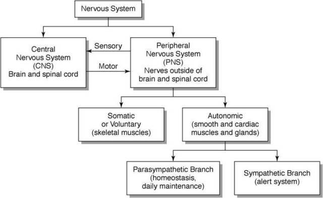
Autonomic nervous system
Autonomic nervous system consists of:
1) The Hypothalamus: which is regarded as the highest control system over the autonomic motor neurons that affect the activity of our visceral organs so the hypothalamus is the center of homeostasis (continuously and unconsciously adjusting the activity of our visceral effectors to match the person’s physical activity and energy requirements).
For example, the heart rate will be adjusted when we are sleeping and or when we are running and this will be done automatically or the heart rate will increase if we are frightened. We can see that hypothalamus is influenced by our emotions that means the hypothalamus is connected to our limbic system. The hypo thalamus is in the area of the brain right above the pituitary gland and we can see that the hypothalamus is connected to the limbic system which is associated with emotions and there are various descending tracts that effects autonomic motor neurons.

Branching of the brain and spinal cord these autonomic motor neurons innervate various internal organs.
There are two types of autonomic motor neurons that innervate each visceral organ of the body (Dual innervation):
1) the parasympathetic autonomic motor neurons that exerts the predominant influence on the visceral organs during the state of relaxation (low energy requirement): rest and digest
2) the sympathetic autonomic motor neurons that exerts the predominant influence on the visceral organs during states of stress (high energy requirement): fight, fright, flight
General patterns of autonomic outflow from the C.N.S
- A) Parasympathetic motor neurons:
The parasympathetic motor neurons are present only in certain cranial nerves and in the sacral spinal nerves: (cranio-sacral)
Cranial nerves III (oculomotor), VII ( Facial), IX ( Glossopharyngeal), X (Vagus) and sacral S2, S3, S4 contains parasympathetic motor neurons.
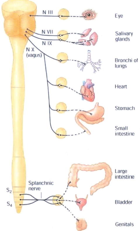
Some of the cranial nerves contain parasympathetic motor neurons and some of the spinal nerves coming out of the sacral level of the spinal cord contains parasympathetic motor neurons. ………
- B) Sympathetic (thoraco-lumbar) division of the Autonomic nervous system:
The only nerves that contain inside them sympathetic motor neurons are those spinal nerves coming of at thoracic and lumbar levels. That’s why the sympathetic nervous system is also known as thoraco-lumbar division. So, there is clear difference anatomically.
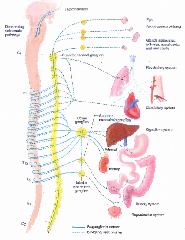
Now what’s interesting is that there are many sympathetic motor neurons that innervate all internal organs instead there is only vagal nerve for parasympathetic nervous system. In other words, all internal organs are innervated by two types of autonomic motor neurons: Dual innervation
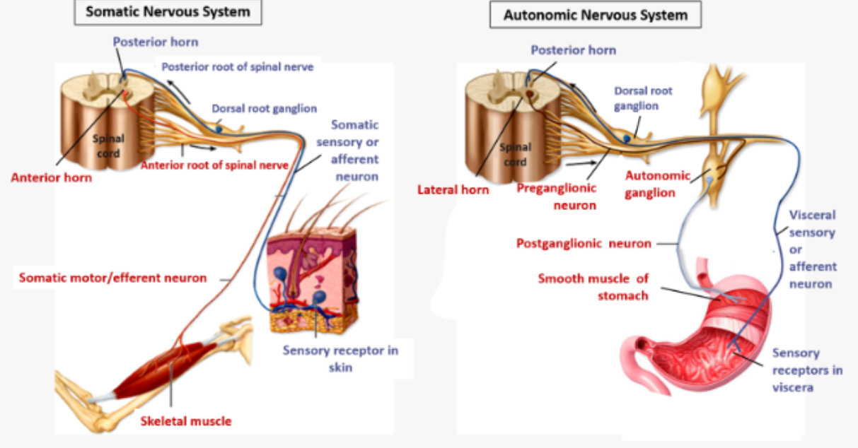
“somatic muscles have nicotinic acetylcholine receptors and are totally dependent of their innervation: they are called neurogenic”
To study the ANS we will take the heart as a simple model (but it could be any other organ)
We know that sympathetic as well as parasympathetic innervation takes two motor neurons to go from the C.N.S to the internal organ. The first neuron is myelinated and the second one is not myelinated (it is the same for Para and sympathetic)
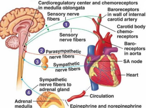
dual innervation of the heart
The acetylcholine is liberated by parasympathetic nerve at the heart (! Acetylcholine receptor at the heart is muscarinic and not nicotinic like the somatic muscles)
Acetylcholine is also released from preganglionic fiber of sympathetic motor neuron as in the preganglionic motor neurons of parasympathetic system but at the level of target organ there is epinephrine release ( we say that post ganglionic fiber is adrenergic)
So in the heart there are two types of receptors ( adrenergic receptors and acetycholine muscarinic receptors)
Somatic muscles havent nicotinic receptors of acetyl choline and if their motor neurons are cut the muscle will be paralysed that means they are totally dependant of the motor neuron ( they are neurogenic) .
Here the dual innervation of the heart is discussed (schematized) but this system can be applied to all of the internal organs of the body .
The adrenergic receptors on the heart are B1 and
Chapter : The Limbic system(anatomy)
The limbic system
The neurologist Paul MacLean has proposed that our skull holds not one brain, but three, each representing a distinct evolutionary stratum that has formed upon the older layer before it, like an archaeological site: He calls it the “triune brain
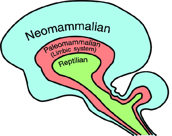 «MacLean, says that three brains operate like “three interconnected biological computers, [each] with its own special intelligence, its own subjectivity, its own sense of time and space and its own memory”. He refers to these three brains as the neocortex or neo-mammalian brain, the limbic or paleo-mammalian system, and the reptilian brain, the brainstem and cerebellum. Each of the three brains is connected by nerves to the other two, but each seems to operate as its own brain system with distinct capacities. This hypothesis has become a very influential paradigm, which has forced a rethink of how the brain functions. It had previously been assumed that the highest level of the brain, the neocortex, dominates the other, lower levels. MacLean has shown that this is not the case, and that the physically lower limbic system, which rules emotions, can hijack the higher mental functions when it needs to.
Reptilian brain:
This brain controls all reflexes, which are automatic and purely regulatory: body temperature, hormonal secretion, blood glucose level, blood pressure, spontaneous respiration…
Ondin’s curse ( lesion or abnormality to the midbrain region: Ondine’s curse—more appropriately known as congenital central hypoventilation syndrome or CCHS—is a rare, severe form of sleep apnea in which an individual completely stops breathing when falling asleep. It is usually congenital, meaning that it is present from birth. It may be noted in the neonatal unit after delivery. Central sleep apnea is characterized by the brainstem failing to prompt normal breathing. This seems to be due to a decreased responsiveness to high levels of carbon dioxide and low oxygen levels within the blood. This becomes especially dangerous during sleep.
Ondine’s curse is named after a mythical tale in which a heartbroken water nymph curses her unfaithful husband to stop breathing should he ever fall asleep. In medical terms, Ondine’s curse represents an extreme form of sleep apnea.
Limbic system (paleo mammalian brain):
Anatomy:
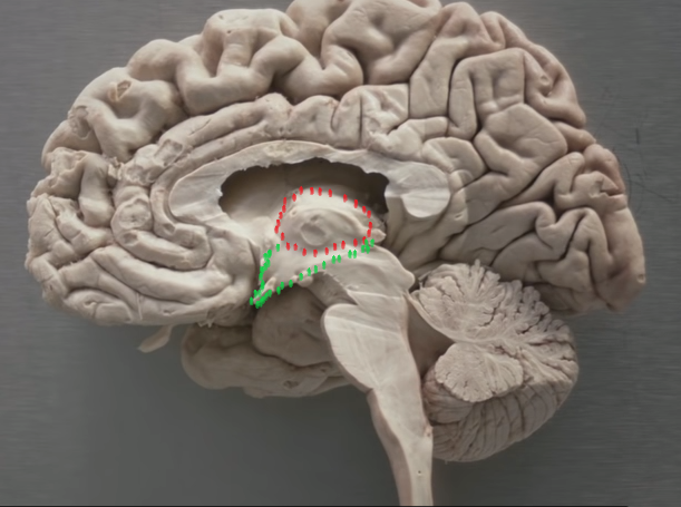
Fig 1: in this mid sagittal dissection the hypothalamus and thalamus are delineated by the hypo thalamic sulcus
Anteriorly the hypothalamus extends to the anterior commissure and the optic chiasm. Inferiorly it includes the mammillary bodies and extends to the infundibular stuck where it communicates with the pituitary gland. The hypothalamus is structurally part of the diencephalon but it functions as part of the limbic system through the reciprocal connections. It helps to maintain homeostasis in the entire body through influences on the endocrine system and importantly through its primary influence on both the sympathetic and parasympathetic systems.
-The limbic system is extremely old from an evolutionary perspective and in its connections; it is interposed between the hypothalamus and the neocortex. Limbic lob is not a true lobe rather it spans the frontal, parietal and temporal lobes, it comprises a ring of cortex in the in the medial surface in the brain (the cingulate gyrus and the para hippocampal gyrus)
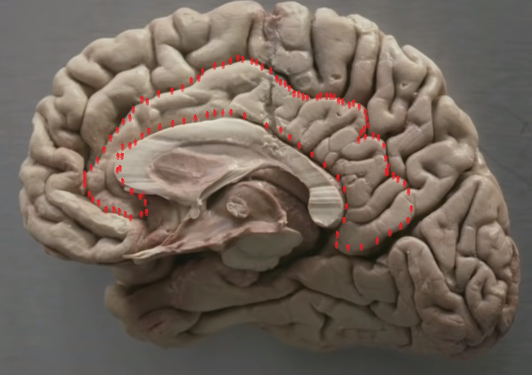

Fig 2 : cingulate gyrus ( left) Parahippocampale gyrus(right)
-The Hippocampus: is primarily involved in memory and it lies in the inferior horn of … And from its posterior ends fibers emerge to form the fornix which swings over the thalamus to rich the mammillary bodies of the hypothalamus by column of the fornix
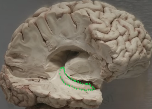
Fig 3 : Hippocampus
– The mammillary bodies are primarily responsible for emotional processing.
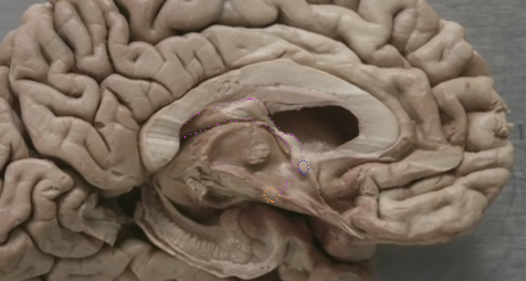
Fig 4 : connection between fornix and mammillary bodies
-The mammillo-thalamic tract connects the mammillary bodies with the anterior nucleus and dorso-medial nucleus of the thalamus and from the thalamus the information travels to the limbic lob (this is the classic papez circuit involved in learning, memory and emotions).
We now know that other structures are involved this circuit including (amygdala…
The amygdala is located in the roof of the inferior horn of lateral ventricle directly underneath the uncus, it lies superior and anterior to the hippocampus (delineated by green dashed cycle)
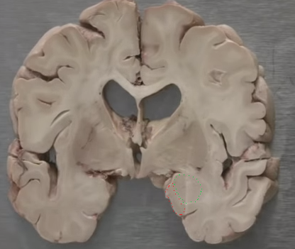
Fig 5 Amygdala (green dashed)
The structure delineated by red is the uncus.The amygdala is a key structure in the expression of emotions, emotional memory and basic drives.