
Fig. 1: Illustration of a Stressful Situation
The image above clearly illustrates a stressful situation triggered by two types of factors:
- The physical stressor
This refers to pain caused by a mechanical injury and detected by mechanical and polymodal nociceptors, as well as free nerve endings.
Within the skin and underlying tissues, there are numerous free nerve endings. Some respond primarily to intense mechanical pressure (mechanical nociceptors), whereas others respond to multiple types of noxious stimuli, including mechanical pressure, extreme heat, and chemical irritation (polymodal nociceptors). These sensory receptors detect tissue injury or potentially damaging stimuli.
Once the alarm signal has been triggered, the electrical signal must travel along the sensory neuron. It is transmitted through two main types of nerve fibers:
- Aδ fibers: relatively large and rapidly conducting fibers that transmit sharp, well-localized, and immediate pain.
- C fibers: thin, slowly conducting fibers that transmit secondary, dull, burning, and more diffuse pain, which may persist after the initial injury.
The signal then travels toward the cell body of the primary sensory neuron, which is located in the dorsal root ganglion (spinal ganglion) (see Fig. 2).
The nociceptive information subsequently enters the spinal cord through the dorsal root, where the primary sensory afferent neurons synapse with second-order neurons, particularly within Rexed laminae I, II, and V. These laminae are anatomically defined layers of the spinal dorsal horn.
Rexed laminae I, II, and V are key regions involved in the reception, processing, and integration of nociceptive signals.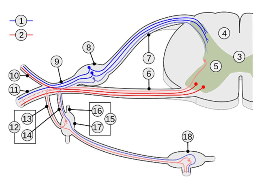 Fig. 2: Neural Mechanisms Underlying the Physical Stress Circuit
The second-order sensory neuron, following synaptic relay within the Rexed laminae of the spinal cord, conveys the nociceptive signal toward higher-order centers. After decussating to the contralateral side of the spinal cord, its axon ascends via the contralateral spinothalamic tract toward supraspinal structures.
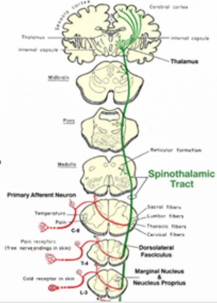 Fig. 3: The Spinothalamic Tract
The spinothalamic tract in the medulla is located in the lateral region, in a dorsolateral position, close to the spinal trigeminal nucleus. It is part of the anterolateral system.
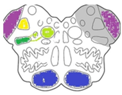 Fig. 4: The Spinothalamic Tract and the Anterolateral System in the Medulla
Bulbar Connections
- Collateral projections to the reticular formation
These projections contribute to the spinoreticular pathway, which is involved in the affective and emotional dimensions of pain, with subsequent projections to the intralaminar nuclei of the thalamus. - Connections with the spinal trigeminal nucleus
These connections contribute to the integration of somatic and facial pain. - Contribution to the spinoparabrachial pathway (indirect)
This pathway provides relay connections to autonomic control centers.
The Spinothalamic Tract in the Pons:
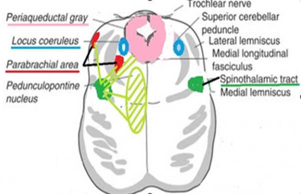
Fig. 5: The Spinothalamic Tract in the Pons
Connections at the pontine level
- Pontine reticular formation: modulation of arousal and participation in autonomic responses to pain.
- Periaqueductal gray (PAG): via ascending projections; an indirect connection that contributes to the initiation of descending pain-modulatory mechanisms.
- Parabrachial complex: projections to the hypothalamus, contributing to autonomic responses and the affective component of pain.
The Spinothalamic Tract in the Midbrain
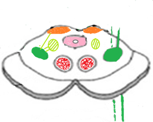 Major Midbrain Connections
- Periaqueductal gray (PAG)
A major center for pain modulation; initiates descending inhibitory pathways and activates the nucleus raphe magnus, leading to spinal inhibition. - Superior colliculus (minor collateral projections)
Involved in orienting reflexes toward nociceptive stimuli. - Midbrain reticular formation
Contributes to arousal and alertness in response to nociceptive stimuli.
Thalamus (Major Relay)
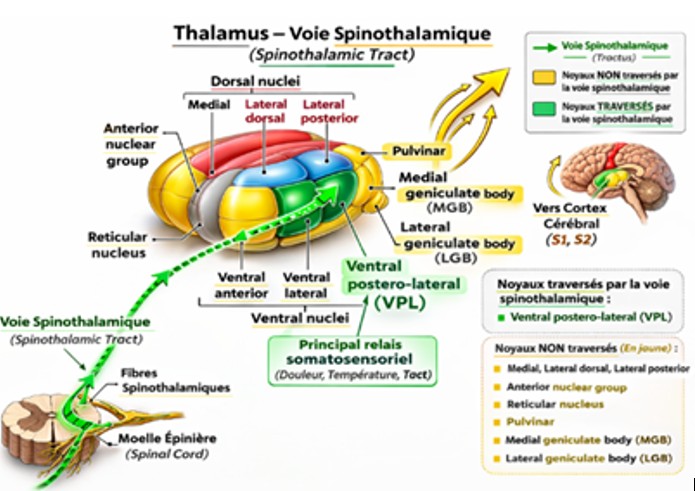
Thalamic Nuclei
- VPL (ventral posterolateral nucleus) → conveys nociceptive information from the body.
- Intralaminar nuclei → involved in the affective and emotional components of pain.
Cerebral Cortex – Conscious Pain Perception
At the cortical level, the primary somatosensory cortex (S1) receives nociceptive information and contributes to the conscious perception of pain, particularly its location and intensity.
Other activated regions:
- Secondary somatosensory cortex (S2) → higher-order processing and integration of somatosensory information.
- Insular cortex → contributes to the subjective awareness of bodily states and the sensory-affective experience of pain.
- Anterior cingulate cortex (ACC) → emotional and affective dimensions of pain, including suffering and distress.
- Amygdala → emotional processing of threat, including fear and defensive responses such as escape or avoidance.
Psychological Stressor: Fear
Fear is a primary survival emotion whose function is to detect potential threats and trigger rapid defensive responses, such as flight, fight, or freezing. In animals such as the gazelle, this response is essential for survival.
Rapid Detection of Threat
Threat detection begins through sensory systems, including:
- Visual system
- Auditory system
- Olfactory system
- Somatosensory system (touch)
The sensory information is then transmitted to the thalamus, which acts as an important relay.
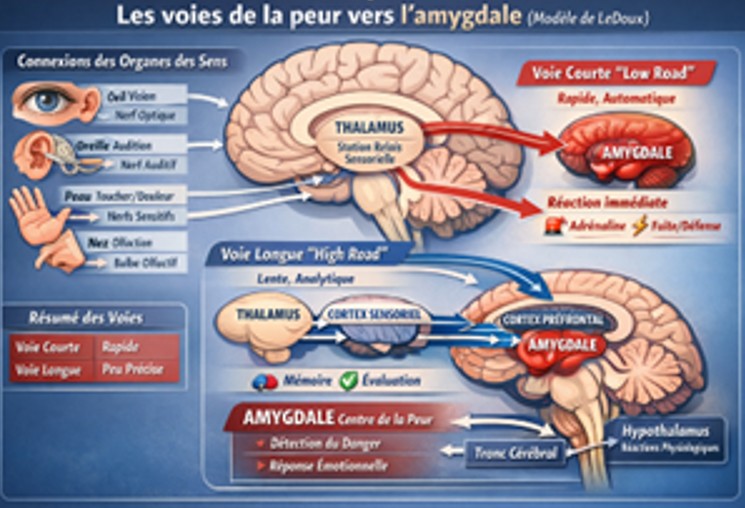
From the thalamus, information can reach the amygdala through two functionally distinct processing routes, as described in the LeDoux model:
-
The fast pathway (“low road”)
Thalamus → AmygdalaThis pathway provides rapid but relatively coarse processing of potential threats. It bypasses detailed cortical analysis and can operate largely outside conscious awareness. Because of its limited sensory resolution, it may generate a false alarm—for example, interpreting a coiled rope as a snake.
-
The slow cortical pathway (“high road”)
Thalamus → Sensory cortex → AmygdalaThis pathway involves more detailed cortical processing before the information reaches the amygdala. It is slower but more precise, allowing conscious evaluation and discrimination of the stimulus—for example, determining whether the apparent “snake” is actually a rope.
1) Response to a Physical Stressor
The information reaches the hypothalamus through the parabrachial nuclei and/or the amygdala, which initiates the fight-or-flight response.
a) Immediate autonomic response (via the hypothalamus and brainstem)
Activation of the sympathetic nervous system:
tachycardia, Increased blood pressure, vasoconstriction of the abdominal and peripheral arteries, vasodilation of the skeletal muscle arteries, mydriasis (pupillary dilation), bronchodilation, sweating, inhibition of gastrointestinal motility, temporary reduction in urine production, as the body prioritizes vital functions such as the heart and skeletal muscles. Reduced renal blood flow leads to decreased glomerular filtration and, consequently, reduced urine output in the short term. In some cases, an urgent need to urinate may occur as a paradoxical response. This constitutes the “fight-or-flight” response.
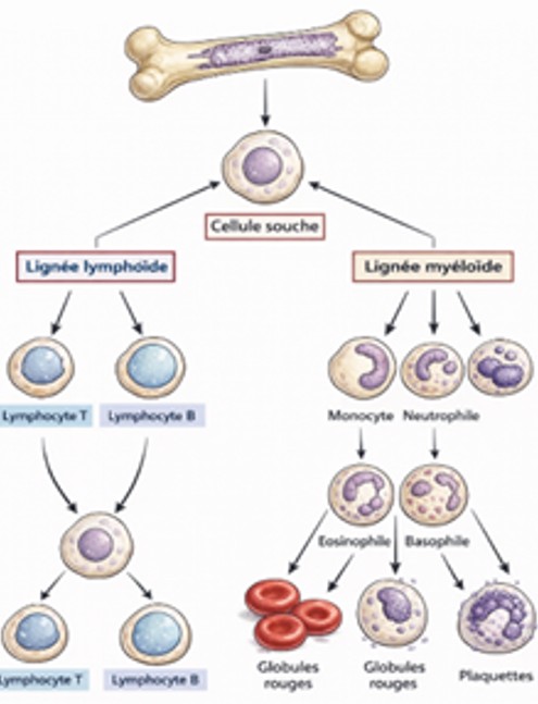
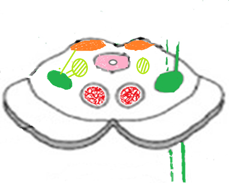
Fig. 8: Stimulation of Bone Marrow Stem Cells
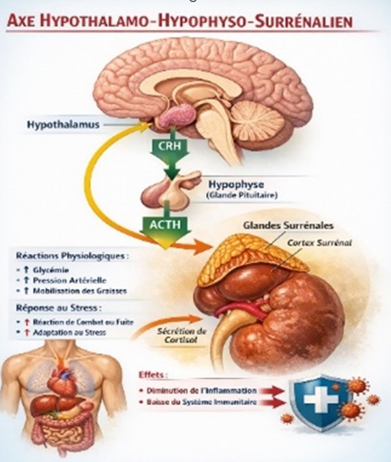
Fig. 9: The Hypothalamic–Pituitary–Adrenal (HPA) Axis
The HPA axis induces the release of cortisol, which exerts an anti-inflammatory effect during the acute stress response.
2) Response to a Psychological Stressor (Fear)
Fear is a primary survival emotion whose function is to detect danger and trigger rapid defensive responses, such as flight, fight, or freezing. In animals such as gazelles, this response is essential for survival.
Rapid detection of danger:
Danger is detected through the sensory systems, including the visual, auditory, olfactory, and somatosensory systems.
The sensory information reaches the thalamus, which acts as a sensory relay and processing center.
Between the thalamus and the amygdala, there are two transmission pathways, as described in the LeDoux model:
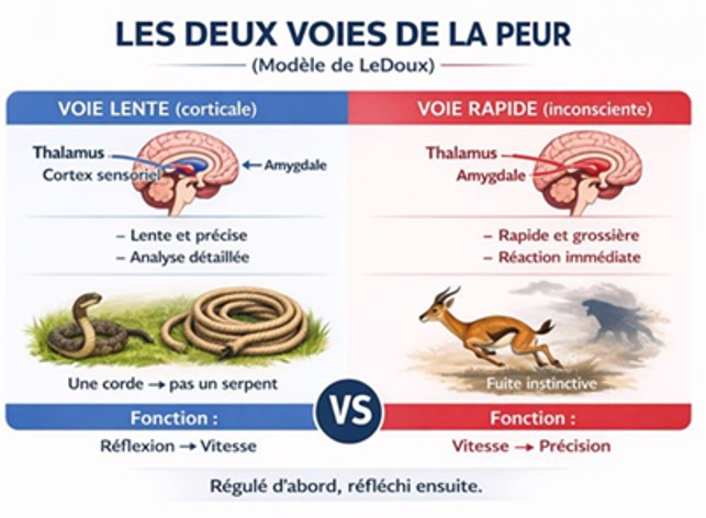
Fig. 10: The Two Fear-Transmission Pathways
En 1937, dans son article scientifique intitulé “A Proposed Mechanism of Emotion”,Papez cherchait à expliquer le fonctionnement des émotions dans le cerveau. Il a proposé un circuit neuronal fermé, aujourd’hui appelé circuit de Papez, reliant plusieurs structures cérébrales.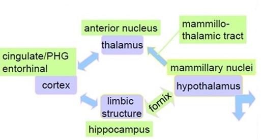
Fig. 11: Papez circuit
Although Papez’s model was subsequently modified, particularly with the introduction of the concept of the limbic system, it remains a major historical foundation in neuroscience. We later realized that this system is far more complex and involves numerous nuclei and interconnected structures, allowing us to experience the extraordinary diversity of human emotions and pleasures.
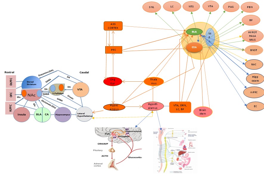
Fig. 12: The Amygdala and Hippocampal Network in the Papez Circuit
In summary, the information reaches all of these nuclei within their respective networks, with the amygdala and hippocampus playing important roles: the amygdala is involved in encoding the emotional value of an experience, whereas the hippocampus records the context and environmental setting associated with the traumatic event. For example, in the case of this gazelle, the entire surrounding environment is unconsciously encoded, whereas the predator itself is consciously perceived and encoded.
The BLA (basolateral amygdala) then stimulates the BNST (bed nucleus of the stria terminalis), which in turn activates the DMH (dorsomedial hypothalamus) and the PVN (paraventricular nucleus of the hypothalamus). The PVN subsequently acts on the sympathetic nervous system and the HPA axis (see above).
The amygdala is also closely interconnected with the prefrontal cortex (PFC), which normally exerts an inhibitory regulatory influence on the amygdala. Under certain conditions, the amygdala becomes hyperactive and is no longer adequately controlled by the PFC. In such circumstances, the stress response becomes dominant, and the patient may experience severe anxiety.
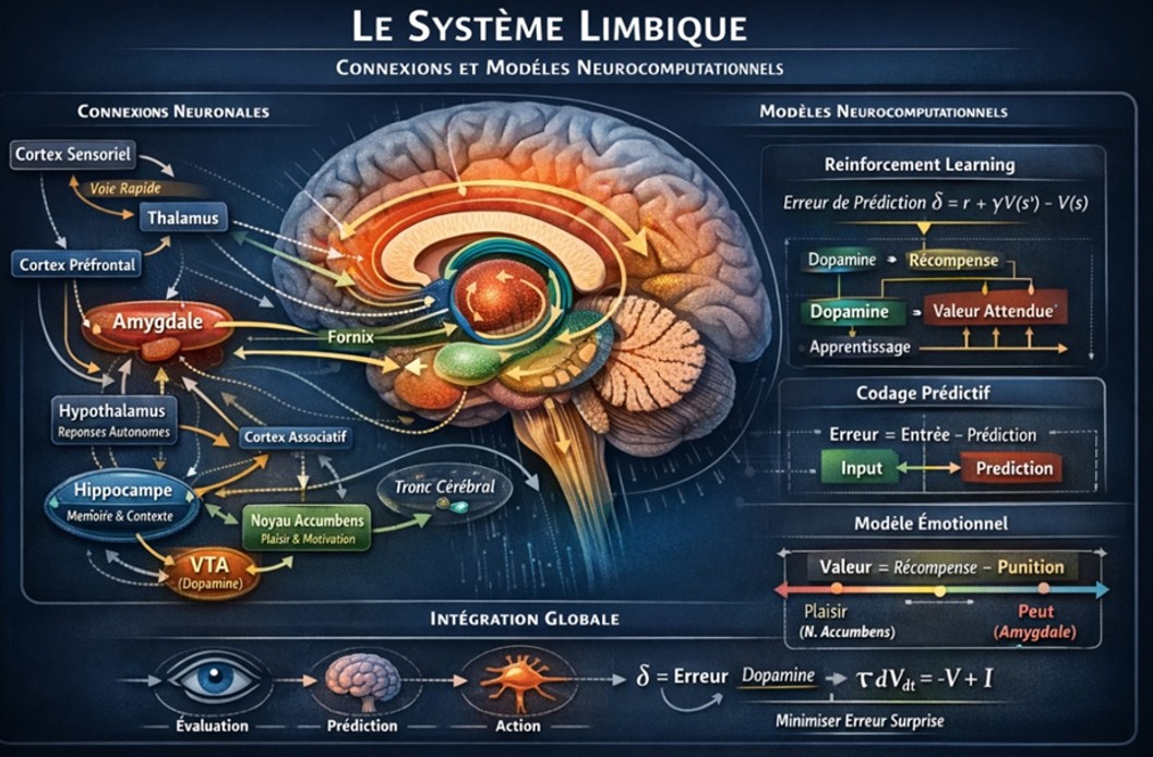
Fig. 13: The Limbic System
2.1 When and How Fear Becomes Traumatic
What is trauma? Trauma occurs when the threat is extreme, pain and fear are intense, a sense of helplessness is present, and the situation overwhelms the individual’s adaptive capacities. In this context, the attack involving claws and the perception of imminent death could potentially be considered traumatic.
In such situations:
- Abnormal encoding of the traumatic memory
- The amygdala becomes hyperactive and over-encodes fear, strongly associating stimuli with danger.
- The hippocampus is inhibited by cortisol, resulting in impaired encoding of the context (time and place), and producing a fragmented, poorly dated memory.
Result: The brain does not properly categorize the event as something that belongs to the past.
1. Failure of prefrontal cortex regulation
Under normal conditions, the prefrontal cortex (PFC) inhibits the amygdala and provides cognitive reassurance, essentially signaling: “It is over; you are safe; calm down.”
Following trauma, prefrontal activity may decrease, leading to a loss of top-down control, while the amygdala remains hyperactive.
Possible manifestations in the gazelle
a) Hypervigilance
- Excessive reactions to the slightest noise
- Inappropriate or disproportionate flight responses
b) Re-experiencing (animal equivalent)
- Reactivation of fear in response to similar stimuli
- Automatic and maladaptive responses
c) Avoidance
- Abandonment of areas that are actually safe
- Changes in feeding behavior
d) Possible freezing response
- Immobility (freeze)
- Failure or inability to initiate an escape response
Key Difference to Remember
| Normal Fear | PTSD |
|---|---|
| Adaptive | Maladaptive |
| Temporary | Persistent |
| Contextualized | Decontextualized |
| Reversible | Self-perpetuating |
Fundamental Message
PTSD is not simply an excess of fear; it is a survival memory that fails to switch off.
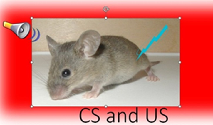
Trauma as Pavlovian Conditioning
Recap: Classical Pavlovian Conditioning
Simple model:
Neutral stimulus (NS) + aversive stimulus (US) → fear response
After repeated pairings or an extremely intense aversive experience, the previously neutral stimulus becomes capable of triggering a fear response on its own.
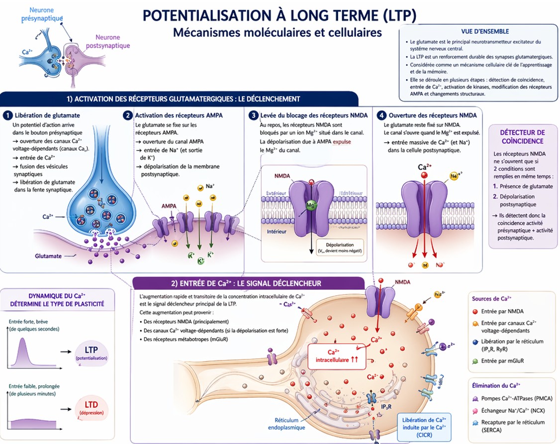
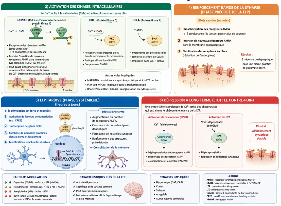
PKA (Protein Kinase A)
 Final Outcome
- Increased number and efficacy of AMPA receptors
- Structural changes, including dendritic spine remodeling
- Long-lasting strengthening of the synapse
Logical Chain Summary
Intense stimulation → AMPA activation → NMDA activation → ↑ Ca²⁺ → activation of CaMKII / PKA / MAPK → ↑ AMPA receptors + structural modifications → LTP (memory)
The Relationship Between Stress and Inflammation
Stress-induced immune responses in the periphery and their underlying mechanisms
Stress-Induced Leukocyte Mobilization from the Bone Marrow
Studies in rodents, primarily using repeated social defeat stress, have shown that stress alters the number, distribution, and properties of leukocytes. In general, chronic stress decreases lymphocytes, such as T cells and B cells, while increasing myeloid cells, such as neutrophils and monocytes, in the blood and bone marrow.
These changes may result from altered leukocyte differentiation and proliferation in the bone marrow, as well as changes in their release into the circulation.
These leukocyte populations do not respond in the same way after cessation of stress. The increase in neutrophils and decrease in B cells persist for several days after the last stress exposure, whereas other leukocyte changes disappear.
Acute stress may induce changes in leukocytes similar to those observed with chronic stress, as a decrease in lymphocytes and an increase in myeloid cells can be detected immediately after a single 2-hour exposure to social defeat stress. However, the effects of acute stress may be less persistent. Twelve hours after a single 2-hour exposure to social defeat stress, only an increase in neutrophils, but not in monocytes, was observed.
These findings indicate that chronic stress induces more persistent leukocyte changes than acute stress, and that the increase in neutrophils persists longer than changes affecting other leukocyte subsets.
Roles of the Autonomic and Neuroendocrine Systems
Stress-induced leukocyte mobilization from the bone marrow is mediated primarily by the autonomic and neuroendocrine systems.
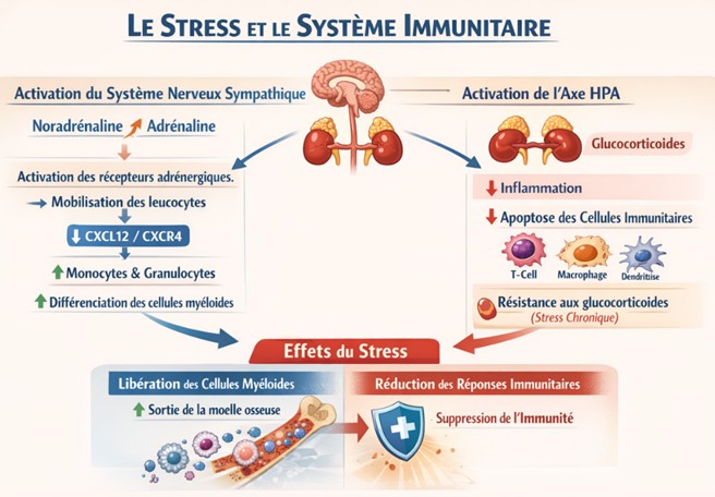
Activation of the sympathetic nervous system (SNS) during stress induces the release of norepinephrine and epinephrine into the blood and tissues. These neurotransmitters activate adrenergic receptors on leukocytes and surrounding cells, such as mesenchymal stem cells in the bone marrow, thereby promoting stress-induced leukocyte mobilization.
Sympathetic activation may be sufficient to produce these effects, as systemic administration of isoproterenol, a non-selective β-adrenergic agonist, decreases lymphocytes and increases monocytes and granulocytes in the bone marrow.
These adrenergic receptors may promote myeloid cell differentiation and proliferation. They may also reduce the expression of C-X-C motif chemokine ligand 12 (CXCL12) by osteoblasts and mesenchymal stem cells. CXCL12 normally contributes to the retention of myeloid cells in the bone marrow through the C-X-C motif chemokine receptor 4 (CXCR4).
In addition, stress activates the hypothalamic–pituitary–adrenal (HPA) axis, increasing circulating glucocorticoid levels.
Glucocorticoids act directly on myeloid and lymphoid cells to suppress inflammatory responses. They also promote the apoptosis of T cells, macrophages, and dendritic cells, thereby inhibiting immune responses.
However, following chronic stress, monocytes and macrophages develop glucocorticoid resistance, or reduced glucocorticoid sensitivity, in both mice and humans. This glucocorticoid resistance can consequently disinhibit inflammatory responses.
At the same time, glucocorticoids can promote innate immune responses to stress by suppressing the CXCL12–CXCR4 pathway, which normally retains myeloid cells in the bone marrow, and by increasing cytokine release from these cells. These effects were prevented by metyrapone, an inhibitor of glucocorticoid synthesis.
Roles of Cytokines and Chemokines
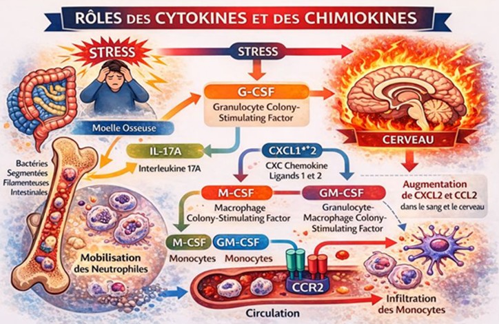
Stress affects the expression of various cytokines and chemokines in the bone marrow and circulation, likely contributing to stress-induced immune responses. Distinct but overlapping cytokine pathways regulate the proliferation and mobilization of neutrophils and monocytes.
G-CSF (Granulocyte Colony-Stimulating Factor) generally promotes neutrophil proliferation and mobilization, although its effects are not exclusively restricted to neutrophils. G-CSF is thought to locally activate the sympathetic nervous system within the bone marrow, thereby promoting neutrophil mobilization. CXCL1/2 and their receptor CXCR2 also stimulate neutrophil mobilization.
M-CSF (Macrophage Colony-Stimulating Factor) and GM-CSF (Granulocyte-Macrophage Colony-Stimulating Factor) promote monocyte proliferation and mobilization. Similarly, CCL2 (C-C motif chemokine ligand 2), also known as monocyte chemoattractant protein-1 (MCP-1), and its receptor CCR2 (C-C chemokine receptor type 2) stimulate monocyte mobilization.
Stress increases circulating levels of G-CSF. Because IL-17 promotes neutrophil mobilization through G-CSF secretion, restraint stress has been shown to increase the number of aged neutrophils through an IL-17A-dependent mechanism, presumably involving intestinal T helper 17 (Th17) cells. Because this neutrophil mobilization depends on segmented filamentous bacteria (SFB) in the gut, IL-17A may provide a mechanistic link between the gut microbiota and the innate immune response to stress.
Indeed, stress alters the gut microbiota, which is associated with both stress susceptibility and stress-related responses. Although the underlying mechanisms remain incompletely understood, the gut microbiota is regulated by multiple pathways, including the vagus nerve, glucocorticoids, leukocytes, and secreted cytokines.
However, the effects of stress on circulating IL-17 levels vary across studies, and it remains to be established whether IL-17A directly mediates stress-induced neutrophil mobilization.
In addition, stress increases circulating and brain levels of CXCL2 and CCL2. The increase in CCL2 is not essential for stress-induced monocyte mobilization into the circulation, since CCR2 deficiency does not prevent this mobilization. In contrast, the increase in CCL2 within the brain is critical for stress-induced monocyte infiltration into the central nervous system.
Effects of Stress on the Immune System Through Multiple Organs
Stress can affect the spleen and lymph nodes, at least in part through sympathetic nervous system projections. Acute stress is thought to induce norepinephrine release from sympathetic nerve terminals in the spleen, which acts on choline acetyltransferase-positive (ChAT⁺) T lymphocytes via β2-adrenergic receptors. These splenic T cells subsequently release acetylcholine, thereby inhibiting the production of pro-inflammatory cytokines by macrophages. This anti-inflammatory pathway is beneficial in various models of inflammatory disease, including ischemic acute kidney injury, and can be activated by vagal pathways connected to the splenic sympathetic nerves.
Chronic stress, such as repeated foot-shock stress or restraint stress, increases the proportion of CD4⁺ T cells exhibiting reduced glycolysis and oxidative phosphorylation, together with increased mitochondrial fission, in the spleen and lymph nodes. This metabolic remodeling appears to contribute to stress-induced anxiety-like behavior.
Chronic stress can also increase splenic plasma cells and enhance antibody-dependent immune responses by activating stress-related brain regions, including the central nucleus of the amygdala (CeA) and the paraventricular nucleus of the hypothalamus (PVN), as well as the splenic nerves. However, this enhancement of humoral responses occurs only under mild stress conditions that do not trigger glucocorticoid release. It therefore does not occur during restraint stress accompanied by substantial glucocorticoid secretion. Thus, humoral immunity is differentially regulated depending on the nature and intensity of the stressor.
Stress can also affect non-immune peripheral organs. Acute stress, for example, may increase IL-6 release from brown adipocytes through direct sympathetic signaling.
Collectively, the interactions among multiple organs—including the bone marrow, spleen, gut, and brown adipose tissue—shape stress-induced innate and adaptive immune responses in a manner that depends strongly on the specific conditions and intensity of stress.
Roles of Bone Marrow-Derived Myeloid Cells
The stress-induced immune responses described above can influence neuronal function and contribute to stress-induced behavioral changes. Multiple barriers separate the brain from the peripheral immune system, and leukocytes are rarely present within the healthy brain parenchyma. Nevertheless, myeloid cells, particularly monocytes, are thought to infiltrate the perivascular spaces of the brain, especially during chronic stress.
Circulating monocytes can adhere to brain vascular endothelial cells and subsequently migrate into the perivascular space. In parallel, an elegant skull transplantation study demonstrated a direct pathway by which bone marrow-derived myeloid cells can migrate from the skull bone marrow to the dura mater, thereby bypassing entry into the circulation.
However, dural myeloid cells would still have to cross additional anatomical barriers—the arachnoid and pia mater—to reach the brain parenchyma.
Repeated social defeat stress increases the number of perivascular monocytes in a CCR2-dependent manner. CCL2, a ligand for CCR2, has been reported to increase in microglia following repeated social defeat stress, suggesting that microglia-derived CCL2 recruits circulating monocytes into the perivascular compartment through CCR2 signaling.
In CCR2-deficient mice, both stress-induced anxiety-like behavior and the associated infiltration of monocytes into the brain are attenuated. These findings suggest that blood-borne monocytes may contribute to the anxiogenic effects of chronic stress.
Repeated social defeat stress also mobilizes neutrophils into the circulation, and this increase persists for a longer period after cessation of stress than the corresponding increase in monocytes. Notably, stress-induced neutrophil mobilization is more pronounced in BALB/c mice than in C57BL/6N mice.
Because BALB/c mice are generally more susceptible to stress than C57BL/6N mice, stress-induced neutrophil mobilization may contribute to genetically determined differences in stress susceptibility.
Role of Calcium (Ca²⁺)
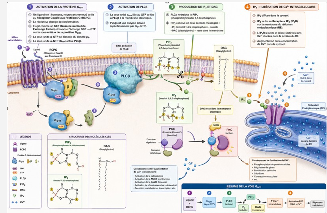
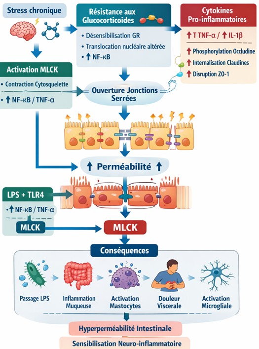
Ca²⁺ binds to calmodulin, leading to the activation of myosin light-chain kinase (MLCK).
Final outcome:
Activation and increased expression of MLCK
3. Function of MLCK
Key reaction:
MLCK catalyzes the phosphorylation of the myosin light chain (MLC):
MLC + ATP → phosphorylated MLC (MLC-P)
Mechanical consequence
Under basal conditions / without stress:
- Low MLC phosphorylation
- Stable actin cytoskeleton
- Intact tight junctions
Under stress conditions:
- ↑ MLC phosphorylation
- Increased contraction of the actin–myosin cytoskeleton
- Mechanical tension is exerted on the tight junctions
- This cytoskeletal contraction pulls on the tight junctions, contributing to their disruption and increased intestinal permeability.
4. Disorganization of Tight Junctions
Normal structure
Key proteins include:
- Claudins
- Occludin
- ZO-1 / ZO-2
These proteins maintain the integrity and sealing of the intestinal epithelial barrier between adjacent cells.
Following MLCK activation
Four major effects:
- Actomyosin contraction
- Dissociation of ZO-1 from the junctional complex
- Internalization of occludin
- Reorganization of claudins
Result:
Opening of the paracellular spaces between epithelial cells
↑ Paracellular permeability
5. Immunological Consequences
Once the intestinal barrier becomes compromised, several luminal molecules can cross the epithelial barrier, including:
- LPS (lipopolysaccharide)
- Peptidoglycans
- Dietary antigens
Immune activation
These microbial products can be recognized by immune cells, including macrophages, through pattern-recognition receptors such as: TLR4 (Toll-like receptor 4)
Cytokine production
This can promote the production of pro-inflammatory cytokines, including:
- TNF-α
- IL-6
- IL-1β
Consequences
Inflammation, which may be:
- Local, within the intestine
- Systemic, when inflammatory mediators enter the circulation
6. Self-Sustaining Feedback Loop — The Vicious Cycle
The system can become self-amplifying:
1. Stress
↓
2. Mast-cell activation
↓
3. ↑ Pro-inflammatory cytokines
4. ↑ MLCK activation/expression
↓
5. ↑ Intestinal permeability
↓
6. Increased translocation of microbial products and luminal antigens
↓
7. ↑ Inflammation
This inflammatory state can further enhance stress responsiveness and perpetuate the cycle.
Contribution of the gut microbiota
- Dysbiosis → disruption of the microbial ecosystem
- ↓ Butyrate → loss of an important intestinal barrier–supporting metabolite
These changes may further aggravate barrier dysfunction and inflammation.
7. Fine Regulation of MLCK
Transcriptional regulation
Transcription factors implicated in the regulation of MLCK expression include:
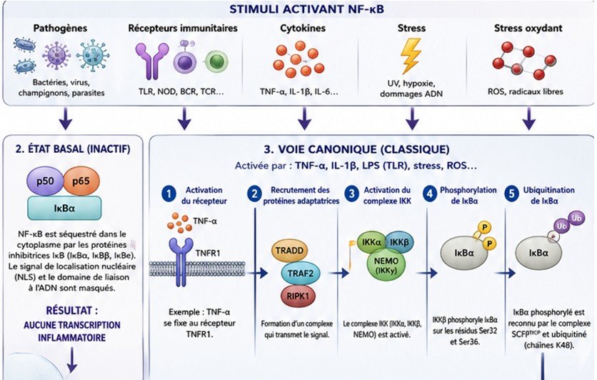
- NF-κB
- AP-1
- C/EBPβ
Post-translational regulation
MLCK activity can also be modulated through:
- Phosphorylation by protein kinases, including PKC and MAPK-related pathways
- Interactions with the cytoskeleton and junctional complexes
Modulators
Potential protective/modulatory factors:
- Butyrate
- Probiotics
- AMPK activation
- Antioxidants, such as N-acetylcysteine
Factors promoting MLCK activation or barrier dysfunction:
- TNF-α
- LPS
- Oxidative stress
- Dysbiosis
8. Experimental Evidence
MLCK inhibition
The MLCK inhibitor ML-7 has been used experimentally to inhibit MLCK activity and can reduce stress- or inflammation-associated increases in intestinal permeability.
Genetic models
Experimental models indicate that:
- MLCK deficiency or inhibition → increased resistance to epithelial hyperpermeability
- MLCK overexpression/activation → increased intestinal permeability
Stress models
Experimental stress paradigms have been associated with:
- ↑ MLCK
- ↑ MLC phosphorylation
- ↑ Intestinal permeability
These effects can be attenuated when MLCK activity is inhibited, supporting a causal role for the MLCK–MLC pathway in epithelial barrier regulation.
Overall Conclusion
MLCK acts as a key biomechanical switch linking extracellular signals to changes in intestinal epithelial barrier function.
It translates:
Psychological and inflammatory signals
↓
Ca²⁺/calmodulin signaling
↓
MLCK activation
↓
MLC phosphorylation
↓
Actomyosin contraction
↓
Tight-junction remodeling
↓
Increased intestinal permeability
Ultra-Synthetic Summary
Stress → CRH + inflammatory signals → ↑ Ca²⁺ → calmodulin → MLCK → MLC phosphorylation → actomyosin contraction → tight-junction opening/remodeling → ↑ intestinal permeability → immune activation/inflammation → vicious cycle
Clinical relevance