Chapter 1: the fundamental units of life
All organisms are made of cells
– The cell is the simplest collection of matter that can live (reproduce)
– Cell structure is correlated to cellular function.
– All cells are related by their descent from earlier cells
Viruses are considered not to live because there reproduction depends on other organisms
How do we study cells?
Normally cells are too small to be seen by eye so we must use micros copes and other techniques like biochemistry to study them
Light microscope : the best magnification 1/1000


The quality of the image depends on 3 things
1) Magnification : rates of object’s image size to its real size
2) Resolution : basically it means the clarity of the image (the minimum size visible)
3) Contrast
Electron microscopy
– Two basic types of electron microscopes are used to study subcellular structures
1) Scanning electron microscopes focus a beam of electrons outs the surface of a specimen, providing images that look there dimensional.
2) Transmission electron microscopes :
Focus a beam of electrons through a specimen (TEMS), there kind of electron microscope is used to study the internal structure of cells.


Example of scanning and transmission electron microscopy


The other technique is Biochemistry
Cell fractionation
– Cell fractionation on takes cells apart and separation the major organelles from one anther
– Ultra centrifuges fractional cells into their component parts.
– Cell fractionation enables scientists to determine the functions of organs
– Biochemistry and cytology help correlate cell function with structure.
Biochemistry
– Cell fractionation takes cells apart and separates the major organelles from the another
– Ultra centrifuges : fractional cells into their component parts
– Cell fractionation enables scientists to determine the functions of organelles
– Biochemistry and cytology helps correlate cell function with structure
Comparing prokaryotic and eukaryotic
– Basic features of all cells
.) Plasma membrane
.) Semifluid substance cells cytosol
.) Chromosomes (carry genes)
.) Ribosomes (make proteins)
– The structural and functional unit of every organism is one of two types of cells : prokaryotic or eukaryotic
– Only organisms of the domains bacteria and arches
– consist of prokaryotic cells
– Protests fungi, animals and plants all consist t of eukaryotic cells.
Prokaryotic cells
– No nucleus
– DNA is an unbound region called the nucleoid
– No membrane bound organ cells
– Cytoplasm bound by the plasma membrane

Eukaryotic cells
– DNA is a nucleus that is bounded by a membranous nuclear envelope
– Cytoplasm in the region between the plasma membrane and nucleus
– Eukaryotic cells are generally much large than prokaryotic cells

In the center of nucleus there is a concentrated region called the nucleolus and this is where ribosomal RNA is made and processed it’s also the site where there’s a lot of organization of other RNA protein complexes that have to be assembled in the nucleus.
And we see the endoplasm reticulum and outside of that is the Golgi and mitochondria and centrosome.
Centrosome is the region where doing cell division the chromosomal DNA comes together and we have also lysosomes, peroxisomes and in the outside of the cell we see microvilli protruding from the cell.
The nucleus :information central
– The nucleus contains most of the cell’s genes and is usually the most consequence…. organelle.
– The nuclear envelope encloses the nucleus, separating it from the cytoplasm
– The nuclear membrane is a double membrane each membrane consists of a lipid bilayer.
– Pores regulate the entry and exit of molecules from the nucleus.
– The shape of the nucleus is maintained by the nuclear lamina which is composed of protein.

Ribosomes: protein factories
– Ribosome are particles made of ribosomal RNA and proteins.
– Ribosomes carry out protein synthesis in two locations:
– In the cytosol (free ribosomes)
– On the outside of the endoplasmic reticulum on the nuclear envelope (band ribosomes)


The endomembrane system regulates protein traffic and performs metabolic functions in the cell.
Ribosome is composed of large submit and small submit and between the 2 submits RNA in the middle and proteins going out in the middle part
The endomembrane system regulates protein traffic and performs metabolic function in the cell.
– Components of the endomembrane system :
– Nuclear envelop
– Endoplasmic reticulum
– Golgi apparatus
– Lysosomes
– Vacuoles
– Plasma membrane
These components are either continuous or connected via transfer by vesicles.
The endoplasmic reticulum: biosynthetic factory
– The endoplasmic reticulum (ER) accounts for more than half of the total membrane in many eukaryotic cells.
– The ER membrane is continuous with the nuclear envelope.
– There are two destinct regions of ER.
.) Smooth ER which lacks ribosomes


.) Rough ER with ribosomes studding its surface.


Functions of smooth ER and rough ER :
The smooth ER:
– Synthesize lipids.
– Metabolizes carbohydrates
– Detoxifies poison
– Stores calcium
The rough ER:
– Has bound ribosomes, which secret glycoproteins (protein covalently bonded to carbohydrates )
– Distributes transport vesicles proteins surrounded by membrane
– Is a membrane factory for the cell
The Golgi apparatus: shipping and receiving center
– The Golgi apparatus consists of flattened membranous sacs called cisternae
– Functions of Golgi apparatus:
.) Modifies products of the ER
.) Manufactures certain macromolecules
.) Sorts and packages molecules into transport vesicles
Lysosomes: digestive compartments
– Some types of cells can engulf other cells by phagocytosis this forms a food vacuole
– A lysosome fuses with the food vacuole and digests the molecules.
– Lysosomes also use enzymes to recycle the cell’ s over organelles and macromolecules a process called autophagy.
Mitochondria and chloroplasts change energy from one form to another.
– Mitochondria are the sites of cellular respiration a metabolic process that generates ATP.
– Chloroplasts, found in plants and algae are the sites of photosynthesis.
– Peroxisomes are oxidative organelles.
– Mitochondria and chloroplasts:
.) are not part of the endomembrane system
.) have a double membrane
.) have proteins made by free ribosomes.
.) contain their own DAN
Mitochondria: chemical energy conversion
– Mitochondria are in nearly all eukaryotic cells
– They have a smooth outer membrane and an inner membrane folded into cristae.
– The inner membrane creates two compartments: inter membrane space and mitochondrial matrix.
– Some metabolic steps of cellular respiration are catalyzed in the mitochondrial matrix
– Cristae present a large surface area for enzymes that synthesize ATP


Chloroplasts: capture of light energy
– The chloroplast is a member of a family of plant organelles called plastids
– Chloroplasts contain the green pigment chlorophyll, as well as enzymes and other molecules that function in photosynthesis
– Chloroplasts are found in leaves and there green organs of plants and in algae.


Peroxisomes: oxidation
– Peroxisomes are specialized metabolic compartments bonded by a single membrane
– Peroxisomes produce hydrogen peroxide and convert it to water.
– Oxygen is used to break down different types of molecules
Chapter 2:the structure of biological membranes
The plasma membrane:Is the boundary that separates, the living cell, from its surrounding and exhibits selective permeability, allowing some substances to cross it more easily than others.
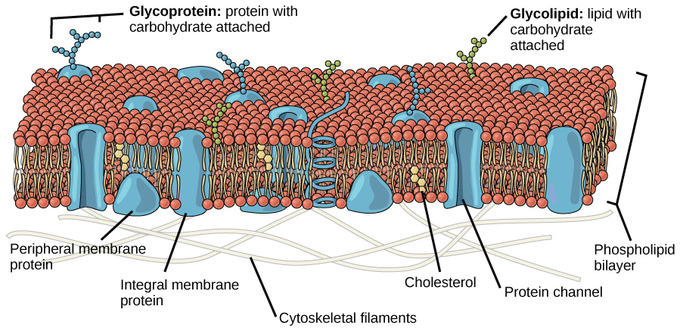
Cellular membranes are fluid mosaics of lipids and proteins
– Phospholipids are the most abundant lipid in the plasma membrane
-Phospholipids are amphipathic molecules, containing hydrophobic and hydrophilic regions.
-The fluid mosaic model states that a membrane is a fluid structure whith a “mosaic” of various proteins embedded in it.
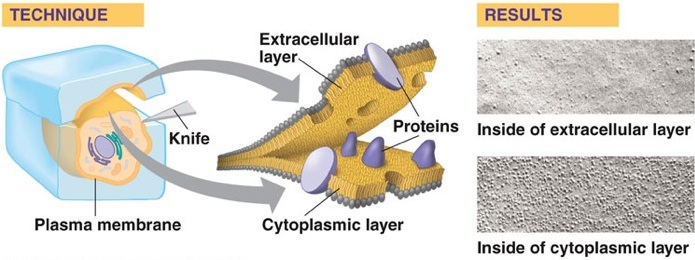
Freeze fracture experiments
– Freeze fracture studies of the plasma membrane supported the fluid mosaic model.
– Freeze fracture is a specialized preparation technique that splits a membrane along the middle of the phosphatide bilayer.
The fluidity of membranes
– Phospholipids in the plasma membrane can move whithin the bilayer
– Most of the lipids and some proteins drift laterally.
– Rarely does a molecule flip-flop transversely across the membrane
Membrane proteins and their functions:
-Peripheral proteins are bound to the surface of the membrane
– Integral proteins penetrate the hydrophobic core
-Integral proteins that span the membrane are called transmembrane proteins.
-The hydrophobic regions of an integral proteins consist of one or more stretches of nonpolar amino acids.
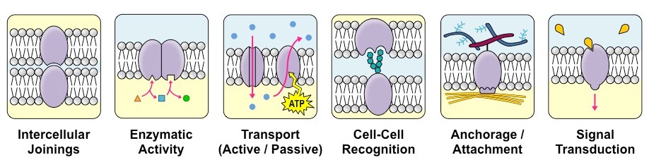
Six major functions of membrane proteins:
– Transport
– Enzymatic activity
– Signal transduction
– Cell-cell recognition
– Intercellular joining
– Attachment to the cytoskeleton and extra cellular matrix
Synthesis and sidedness of membranes
– Membranes have a distinct inside and outside faces
-The asymmetrical distribution of proteins, lipids and associated carbohydrates in the plasma membrane is determined when the membrane is built by the ER and Golgi apparatus.
Membrane structure results in selective permeability
– A cell must exchange materials with its surrounding, a process controlled by the plasma membrane
– Plasma membranes are selectively permeable, regulating the cell’s molecular traffic
– Hydrophobic (nonpolar) molecules such as hydrocarbons, can dissolve in the lipid bilayer and pass through the membrane rapidly
– Polar molecules such as sugars do not cross the membrane easily
Passive transport: diffusion across a membrane with no energy investment
1)Diffusion is the tendency for molecules to speed out evenly into the available space.
2)Although each molecule moves randomly, diffusion of a population of molecules may exhibit a net movement in one direction
3) At dynamic equilibrium, as many molecules cross one way as cross in the other direction.
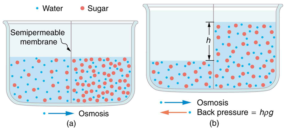
Effects of osmosis on water balance
1) Osmosis is the diffusions of water across a selectively permeable membrane.
2)Water diffuses across a membrane from the region of lower solute concentration to the region of higher solute concentration.
Water balance of cells without walls. Tonicity is the ability of a solution to cause a cell to gain or lose water.
Isotonic solution: solute concentration is the same as that inside the cell, no net water movement across the plasma membrane
Hypertonic solution: solute concentrations greater than that inside the cell; cell loses water
Hypotonic solution: solute concentration is less than that inside the cell; cell gains water.
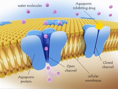
Transport proteins: Transport proteins allow passage of hydrophilic substances across the membrane.
Some transport proteins, called channel proteins have a hydrophilic channel that certain molecules or ions can use as a tunnel.
Channel proteins called Aquaporin facilitate the passage of water.
Facilitated diffusion, passive transport aided by proteins, facilitated diffusion : transport proteins speed the passive movement of molecules across the plasma membrane.
Channel proteins provide corridors that allow a specific molecule or ion to cross the membrane
Channel proteins include:
Aquaporin, to facilitate diffusion of water ,Ion channels that open or close in response to a stimulus (gated channels)
Active transport uses energy to move solutes against their gradients:Facilitate diffusion is still passive because the solute moves down its concentration gradient.Some transport proteins however can move solutes against their concentration gradient.Active transport moves substances against their concentration gradient.Active transport requires energy usually in the form of ATP.Active transport is performed by specific proteins embedded in the membranes.
Ion pumps maintain membrane potential:Membrane potential is the voltage difference across a membrane. Voltage is created by differences in the distributions of positive and negative ions.
Two combined forces collectively called the electrochemical gradient, drive diffusion of ions across a membrane. A chemical force (the ion’s content ratio gradient),An electrical force (the effect of the membrane potential on the ion’s movement) An electro genic pump is a transport protein that generates voltage across a membrane. The sodium potassium pump is the major electro genic pump of animal cells.The main electro genic pump of plants, fungi and bacteria is a proton pump.µ
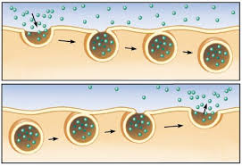
Bulk transport by exocytosis and endocytosis
Small molecules and water enter or leave the cell through the lipid bilayer or by transport proteins
Large molecules, such as polysaccharides and proteins, cross the membrane in bulk via vesicles.
Bulk transport requires energy.
In exocytosis transport vesicles migrate to the membrane, fuse with it and release their contents.
In endocytosis the cell takes in macromolecules by forming vesicles from the plasma membrane.
Chapter 3: eukaryote cell division
Cell division is incredibly tightly regulated (cancer is described as an unregulated cell growth).In respect tothe cell division,there is a machinery that is a transitory state and this machinery is renewed every cell cycle.
Key components of this machinery are:
1) Spindle: is made of microtubules or cytoskeleton, centrioles, kinetochores and contractile ring.
2) And this machinery is controlled by a control system which integrates signals from growth and division.
These signals are:
a) Cell division kinases (CDK)
b) Each kinase has an activity that is controlled by a cycline (regulatory subunit of CDK) and the activity
of these 2 systems is controlled by a third system which is;
c) regulated destruction mechanism.

Cell division is highly regulated:
The cells are dividing by 8 minutes (but neurons by 100 years) but the typical cells (eg liver cells) divides
1/year, and the cells of gut are dividing 1/day .
Cell division
1) Cell division requires partitioning of organelles for this there is 3 stratégies :
A) Abundant organelles like ribosomes peroxisomes ( 1/2 goes in one side and 1/2 to the other side)
B) Mitochondria chloroplast: these are ancient captured bacterias and conserves their own mechanism
of division .So chloroplast and mitochondria split during cell division .
C) Golgi and endoplastic reticulum becomes small vesicles and then after cell division they come back
together and reform Golgi and ER
D) Centrosome = centriole is the poorly understood in eukaryotes cells.
Centriole template his own replication so we need a centriole to make a new centriole (centriole play a key
role in mitotic spindle apparatus and centrioles are structures, that DNA contemplate his replication.


G1 and G0: G1 is the phase in which the cell ask herself “am I big enough to divide” are there enough food?
There are also the + or – factors from other cells or the environment.And if the final decision is not
to divide the cell goes back to G0 phase where there is no division.


S phase : the phase in which nuclear DNA replication and mitochondrial DNA replication occurs .
G2: another decision making phase is G2: cell verify if every single genome has been replicated or not. If it has not, there is a mechanism that ask the cell to halt in G2 until the last nucleotide could be replicated. G2 will be prolonged until the cell has all needed to go to M.Another control point is to see if each kinetochore archived bipolar spindle attachment.
M: and finally mitosis happens:
Chromosomes consist of one DNA molecule and the proteins associated with that and we must not underestimate the importance of the proteins and the mass of proteins associated with the DNA (their mass exceeds the mass of DNA).
There are critical landmarks on the chromosomes and the most important is the centromere. The centromere is the point at which the chromosome attaches to the spindle apparatus to allow chromosome segregation that is it’s only function; but at this point there is a very complete protein structure (fifty different proteins called connect cord. So, the connect cord is the structure that assembles at this point to archive protein segregation).
We know that there are the origin of replication on DNA, result in eukaryote cells there are multiple origins of replication.
At the very end of chromosomes, there are specialized structures called telomerase.
Telomerase the enzyme which intervene in the replication of telomere.
Chromosomes have lots of genes collectively our chromosomes.
How 22250 genes which make proteins as well as RNA
Ploidy = number of sets of chromosomes
Haploid = organisms who have one set of chromosomes
Diploid = organisms who have two sets of chromosomes
Triploid = organism who have three sets of chromosomes
Tetraploid = organism who have four sets of chromosomes
Hexaploid = organism who have six sets of chromosomes
Octaploid = organism who have eight sets of chromosomes
The letter N is used to refer to number of chromosome haploid cell.
So, for humans and potatoes N=23 and as we are diploid, so we have 2 x 23 chromosomes
C value refers to the contact of DNA (amount of DNA in the cell)
N is a discontinuous functions but the C value is the continuous function.
So, the amount of DNA in a diploid nucleons is 2 C in G1 and the amount of C in G2=4C
But in G2 the number of chromosomes dose not change.
How do we measure DNA content?
We can do that because of amazing machine
FACS= fluorescent activated cell sorter or cell scanner
The fluorescent dye that steins DNA but it stains DNA proportionately to the DNA reserve