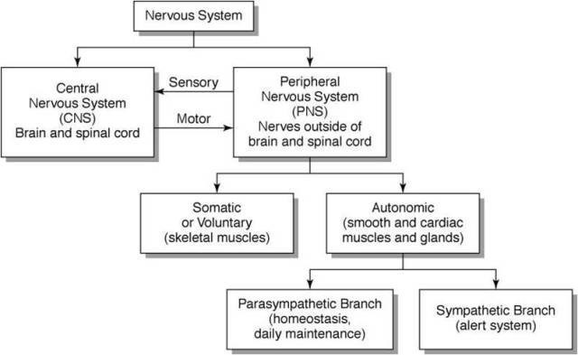
Autonomic nervous system consists of:
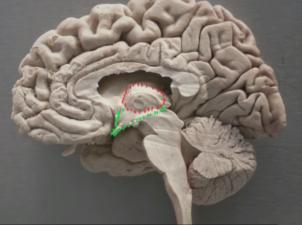
1) The Hypothalamus: which is regarded as the highest control system over the autonomic motor neurons that affect the activity of our visceral organs so the hypothalamus is the center of homeostasis (continuously and unconsciously adjusting the activity of our visceral effectors to match the person’s physical activity and energy requirements).
For example, the heart rate will be adjusted when we are sleeping and or when we are running and this will be done automatically or the heart rate will increase if we are frightened. We can see that hypothalamus is influenced by our emotions that means the hypothalamus is connected to our limbic system. The hypo thalamus is in the area of the brain right above the pituitary gland and we can see that the hypothalamus is connected to the limbic system which is associated with emotions and there are various descending tracts that effects autonomic motor neurons.
Branching of the brain and spinal cord these autonomic motor neurons innervate various internal organs.
There are two types of autonomic motor neurons that innervate each visceral organ of the body (Dual innervation):
1) the parasympathetic autonomic motor neurons that exerts the predominant influence on the visceral organs during the state of relaxation (low energy requirement): rest and digest
2) the sympathetic autonomic motor neurons that exerts the predominant influence on the visceral organs during states of stress (high energy requirement): fight, fright, flight
General patterns of autonomic outflow from the C.N.S
- A) Parasympathetic motor neurons:
The parasympathetic motor neurons are present only in certain cranial nerves and in the sacral spinal nerves: (cranio-sacral)
Cranial nerves III (oculomotor), VII ( Facial), IX ( Glossopharyngeal), X (Vagus) and sacral S2, S3, S4 contains parasympathetic motor neurons.
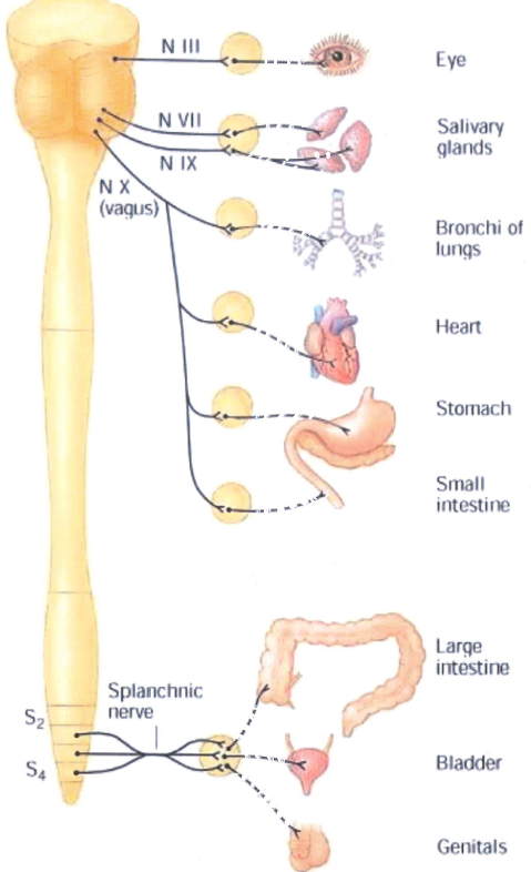
Some of the cranial nerves contain parasympathetic motor neurons and some of the spinal nerves coming out of the sacral level of the spinal cord contains parasympathetic motor neurons. ………
- B) Sympathetic (thoraco-lumbar) division of the Autonomic nervous system:
The only nerves that contain inside them sympathetic motor neurons are those spinal nerves coming of at thoracic and lumbar levels. That’s why the sympathetic nervous system is also known as thoraco-lumbar division. So, there is clear difference anatomically.
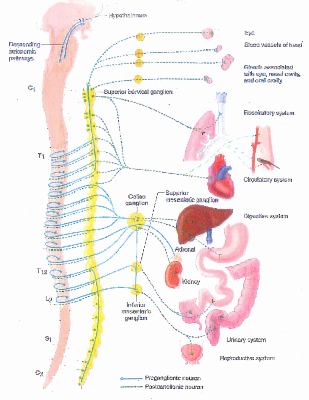
Now what’s interesting is that there are many sympathetic motor neurons that innervate all internal organs instead there is only vagal nerve for parasympathetic nervous system. In other words, all internal organs are innervated by two types of autonomic motor neurons: Dual innervation
“somatic muscles have nicotinic acetylcholine receptors and are totally dependent of their innervation: they are called neurogenic”
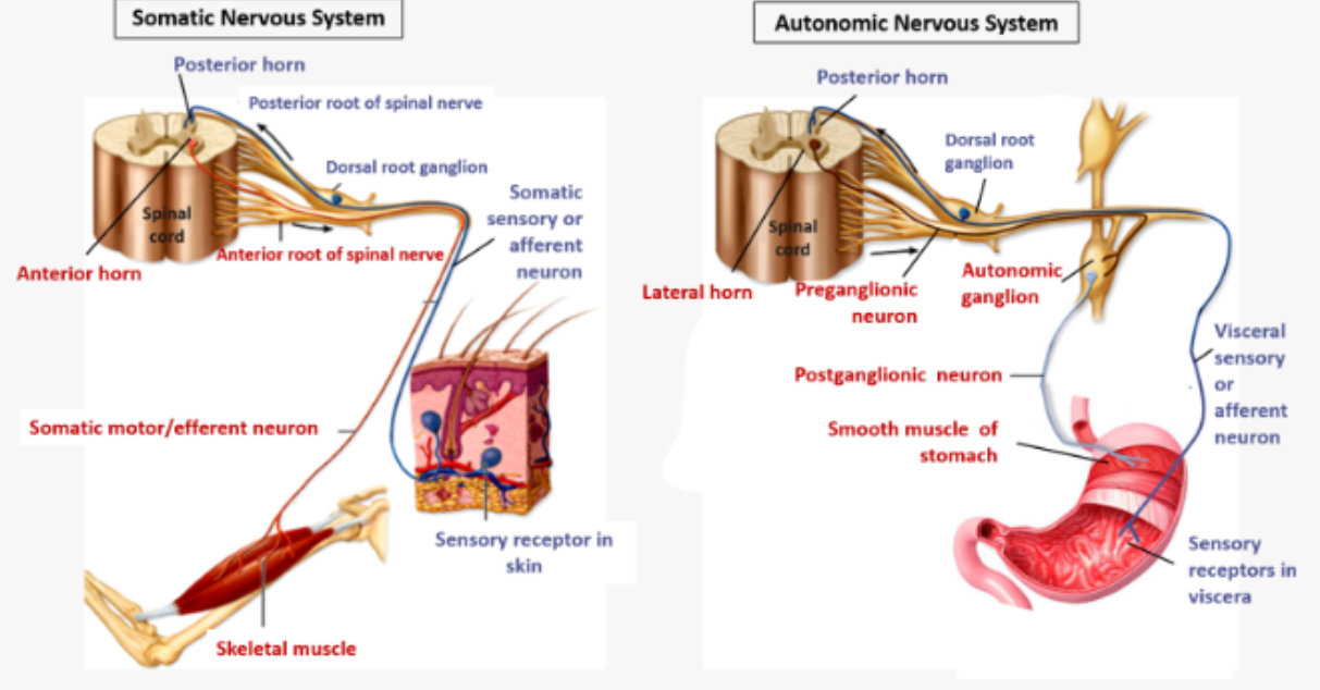
To study the ANS we will take the heart as a simple model (but it could be any other organ)
We know that sympathetic as well as parasympathetic innervation takes two motor neurons to go from the C.N.S to the internal organ. The first neuron is myelinated and the second one is not myelinated (it is the same for Para and sympathetic)
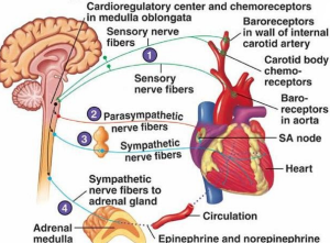
dual innervation of the heart
The acetylcholine is liberated by parasympathetic nerve at the heart (! Acetylcholine receptor at the heart is muscarinic and not nicotinic like the somatic muscles)
Acetylcholine is also released from preganglionic fiber of sympathetic motor neuron as in the preganglionic motor neurons of parasympathetic system but at the level of target organ there is epinephrine release ( we say that post ganglionic fiber is adrenergic)
So in the heart there are two types of receptors ( adrenergic receptors and acetycholine muscarinic receptors)
Somatic muscles havent nicotinic receptors of acetyl choline and if their motor neurons are cut the muscle will be paralysed that means they are totally dependant of the motor neuron ( they are neurogenic) .
Here the dual innervation of the heart is discussed (schematized) but this system can be applied to all of the internal organs of the body .
The adrenergic receptors on the heart are B1 and
